Molecular mechanisms of centriole distal appendage assembly
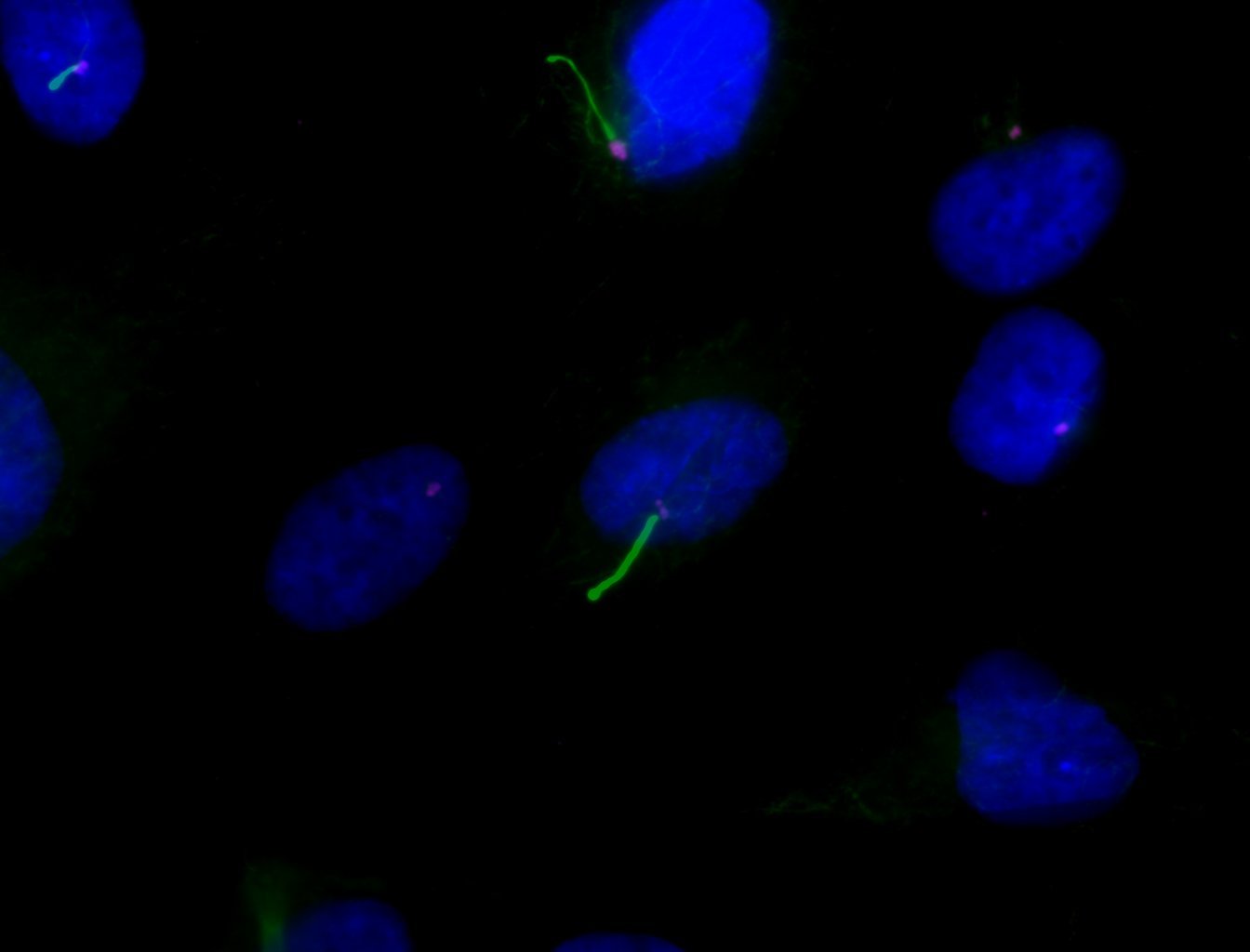
Centrioles are beautiful microtubule-based organelles arranged in a 9-fold symmetry. Mother centrioles contain electron-dense structures called distal appendages (DAPs) that support centriole docking during the onset of cilia formation. We are interested in understanding how distal appendages are assembled, how proteins are recruited to DAPs, and whether some proteins are transient passengers or more stable resident/structural components. In the last few years, great efforts have been made to improve our understanding of how distal appendage proteins are organized and recruited to centrioles. We are actively involved in these efforts.
How do centriole distal appendage proteins regulate ciliogenesis
We have shown that centriole distal appendages mediate centriole-to-membrane docking to initiate ciliogenesis. We are now interested in understanding how this process is regulated, and in mapping out the signaling pathways that regulate these steps, and any post-translation modifications associated with this regulation (e.g. phosphorylation events). These molecular events are linked to how proteins are recruited to distal appendages, and to how different trafficking events regulate the recruitment of the ciliary vesicle and cilia initiation. Our work on the regulation of ciliogenesis by distal appendages has proven to be important in understanding the molecular aetiology of unknown ciliopathies.
Centriole signal transduction pathways
Signal transduction events are fascinating action/reaction steps that regulate biological outcomes in the cell both in health and disease. Centrioles and cilia are characterized by hosting unique signalling molecules and trafficking events. Even within a centrosome, each of the two centrioles is different, which results in one of them being able to function as a basal body and support ciliogenesis. We are interested in understanding the unique signals generated by centrioles and cilia. This is a relatively young field that is only now beginning to develop. These are exciting times!
Cilia Signalling in Polycystic Kidney Disease
Cilia are present in most cells in our body. Thus, it is not surprising that cilia defects can result in many illnesses. These disorders are collectively known as ciliopathies. Many ciliopathies are characterized by kidney defects. However, the role of cilia in PKD is unclear. Although intact cilia are necessary for a cilia-dependent cyst activation (CDCA) pathway, which is normally suppressed by polycystins, a lack of cilia has also been shown to promote cystic phenotypes. Therefore, the exact contribution of ciliary signals to cyst formation remains ill-defined. We want to understand how ciliary signalling promotes PKD to identify potential therapeutic targets that can be taken to the clinic.
Cilia Signalling in Cancer
Cilia are sensory organelles that detect mechanical and chemical stimuli from the environment. Some of these signals can function as pro-survival cues, as exemplified by the Hedgehog pathway. Cilia have been shown to both promote or suppress tumorigenesis, depending on the type of oncogenic lesion that drives tumorigenesis. We are interested in understanding how cilia contribute to cancer. We have shown that primary cilia mediate diverse kinase inhibitor resistance mechanisms in cancer. We are continuing with our efforts to flesh out the molecular mechanisms of cilia-driven drug resistance and to understand how centrioles and cilia support oncogenic phenotypes such as cell invasion.


